The selection of an appropriate spinal screw for each surgical case requires integrating patient anatomy, bone quality, surgical approach, and the biomechanical demands of the specific spinal construct being built. No single screw specification suits every case — the experienced spine surgeon matches screw design, diameter, length, and head type to the individual patient's requirements in each procedure.
Pedicle screw diameter selection is primarily governed by the diameter of the pedicle being instrumented, measured on preoperative CT imaging. The general principle is to select a screw whose outer diameter fills 70 to 80% of the pedicle's inner cortical diameter, maximising cortical contact and pull-out resistance while maintaining a safety margin against cortical breech. In practice, lumbar pedicle diameters vary from 6mm to over 14mm between patients and between spinal levels, requiring a screw range from 4.5mm to 8.5mm outer diameter to cover routine lumbar spine surgery. Thoracic pedicles are substantially smaller — often 3 to 6mm — requiring correspondingly smaller screws that maintain sufficient shaft strength for the forces applied in thoracic deformity correction constructs.
Screw length determines how deep into the vertebral body the screw penetrates beyond the pedicle, and consequently the amount of cancellous bone engaged in the fixation. Standard teaching recommends purchasing 70 to 80% of the vertebral body depth to maximise pull-out resistance while maintaining a safety margin from the anterior cortex and adjacent vascular structures. In osteoporotic bone, longer screws that approach the anterior cortex maximise available cancellous bone engagement, reducing the risk of screw loosening through the low-density trabecular matrix. Preoperative measurement from CT or lateral fluoroscopic images enables accurate screw length planning — empirical selection without measurement increases the risk of either inadequate purchase or cortical perforation.
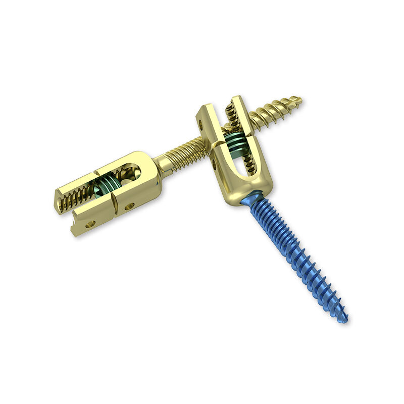
Reduction Pedicle Screw III — Advanced Spinal Fixation — Youbetter Medical (youbettermed.com)
Standard pedicle screws have fixed-height tulip heads that accept the connecting rod at the anatomical level of the screw head. In spondylolisthesis — where one vertebra has slipped forward on the one below — a standard screw in the slipped vertebra will be significantly anteriorly displaced relative to the adjacent level, making rod seating technically difficult and corrective reduction forces difficult to apply. Reduction pedicle screws address this with extended, telescoping tulip heads that can be set at a height above the screw head before the rod is seated, then driven down onto the rod to translate the slipped vertebra posteriorly into a reduced position. The Reduction Pedicle Screw III in Youbetter Medical's spine system provides this reduction capability with a robust, reliable reduction mechanism designed for the significant forces required in spondylolisthesis correction.
Cervical pedicle screws offer superior fixation strength compared with lateral mass screws — the conventional alternative for posterior cervical instrumentation — but require substantially more precise placement because of the narrow pedicle dimensions and the proximity of the vertebral artery medially and the nerve root inferolaterally. Only surgeons with specific experience in cervical pedicle screw placement, supported by CT navigation or intraoperative CT confirmation of screw trajectory, should employ cervical pedicle screws in clinical practice. Youbetter Medical's cervical pedicle screw system is designed for these demanding precision requirements, with diameter options appropriate for the smaller cervical pedicle dimensions and full compatibility with their cervical spine instrument sets.